TAKANO LAB
LABORATORY FOR DYNAMIC IMAGING OF NERVOUS SYSTEM AND FUNCTION

Our Work
Our research aims to understand the circuit mechanisms of epilepsy. We use a rodent model of epilepsy and apply various cutting-edge technologies such as two-photon microscopy, optogenetics, microelectrode arrays, and virtual reality navigation.
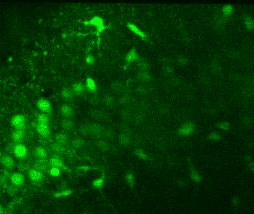
in vitro dynamic cellular imaging
Using in vitro dynamic cellular imaging such as calcium and chloride imaging, we study cellular and circuit mechanisms of seizure initiation and termination in models of temporal lobe epilepsy.
in vivo dynamic cellular imaging
We integrated a virtual reality (VR) navigation system with an in vivo two-photon microscopy setup to conduct multi-cellular calcium imaging of hippocampal neurons while a mouse navigates a VR linear track. We develop image and data analysis algorithms to analyze calcium transients and place cells.


Opto-IHK Model
By combining the intrahippocampal kainate (IHK) model of TLE with optogenetic stimulation of hippocampal neurons, we demonstrated that a brief illumination of blue light can trigger convulsive seizures that are electrographically and behaviorally identical to spontaneous seizures. We have shown that this method can rapidly screen anti-epileptic drugs. (Chen et. al., 2024)
Graphene Microelectrode Array
We used graphene microelectrode technology to monitor acute cortical seizures (Driscoll et al., 2021). We developed a cannula graphene assembly for recording and imaging seizures in deeper brain structures (Mulcahey et al., 2022).
